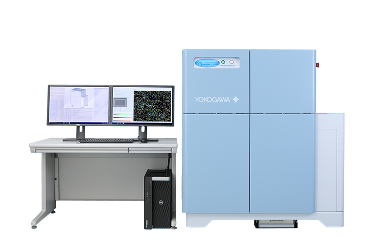
Yokogawa CV8000 Confocal Imaging Platform
The Best High Content Confocal Screening
Wako Automation is the exclusive European distributor of the Yokogawa CV8000
Yokogawa imaging systems provide expanded flexibility by enabling confocal and live cell assays with sufficient signal and a biologically relevant imaging period. Explore confocal imaging of live cells in fast time-lapse mode at up to 49fps or long-term confocal time lapse imaging of cells over multiple days. Confocal imaging on Yokogawa systems is the gold standard powering labs of the future.
Yokogawa’s line of High Content Imaging and Analysis products utilize the CSU-W1 confocal spinning disk and Hamamatsu’s ORCA Flash 4.0 V3 sCMOS cameras. One system with the latest, most powerful confocal spinning disk combined with the most sensitive sCMOS camera available today.
Follow up with High Content Analysis using Yokogawa’s Cell Pathfinder (CPf) analysis software and Wako Automation’s Wako Software Suite (WSS) designed to quickly search for objects of interest in low resolution mode and quickly re-image in high resolution.
Overview
The Yokogawa CV8000 is the fastest, highest resolution, and most flexible High Content confocal solution for today’s scientist. The CV8000 is also the ideal imaging system for live cell multi-day assays, tissue section imaging on slides, performing high speed kinetic imaging, imaging Cell Painting Assays, and spheroid/organoid imaging. Yokogawa has continuously improved its systems culminating in the unrivaled CV8000 which combines the highest resolution spinning disk available today, the CSU-W1, with the highest sensitivity sCMOS camera, the Hamamatsu ORCA Flash 4.0 V3.
The CV8000’s flexible design enables scientists to visualize small objects more clearly because of the greater resolution provided by the latest Yokogawa confocal unit and the amazing quantum efficiency of the best sCMOS camera on the market. In addition, resolution and light gathering can be further improved by purchasing Yokogawa’s optional 20x, 40x, and 60x water immersion objectives.
Are you imaging spheroids, organoids or large objects and need to improve speed and image quality? The optional dual 25µm and 50µm confocal pinhole option further improves resolution beyond anything else available for HCS. The 20x 1.0NA water objective combined with the 25µm pinhole enables high quality spheroid images, reduces or eliminates the need to stitch dozens of images together, and provides higher quality results and a speedier workflow.
CV8000 Features
Microscopists have trusted the Yokogawa name since they developed the first microlens enhanced Nipkow spinning disk almost 25 years ago. Microscopy is at the heart of High Content Imaging. Trust the preferred brand of microscopists worldwide.
- The CV8000 provides up to four large format Hamamatsu Orca Flash 4.0 V3 sCMOS cameras with simultaneous acquisition in up to four channels for unrivaled speed. The CV8000 beats most systems under most conditions even when using sequential imaging. Sequential imaging eliminates 100% of crosstalk that can occur from simultaneous imaging.
- Yokogawa’s 60x, 40x, and 20x high NA water immersion objectives capture more light and provide high image resolution, which is especially helpful for spheroid and tissue imaging.
- Image in fluorescence, bright field, true phase contrast, or digital phase contrast modes to see which mode meets your imaging needs.
- The sCMOS sensor provides almost 4x larger field of view compared to traditional CCD sensors allowing scientists to image more cells per field.
- The CV8000 uses Hamamatsu’s cutting-edge ORCA flash 4.0 V3 sCMOS sensor providing the highest quantum efficiency of any sCMOS on the market today. This high sensitivity camera enables CV8000 users to see objects not possible on other microscopes. Increasing exposure times also increases background. Nothing can make up for a camera with a low quantum efficiency.
- Flexibility is key with the CV8000. The 25µm pinhole option provides another level of resolution and enables the use of lower magnification objectives without sacrificing image quality. Use the 20x water objective with the new 25µm pinhole option and make image stitching a thing of the past.
- Yokogawa invented the microlens enhanced confocal spinning disk to enable confocal imaging of live samples while significantly reducing the risks of phototoxicity and bleaching. The CV8000 provides temperature, humidity, and CO2 control to enable true live cell multiday imaging.
- The optional FRET mode further expands your assays choices.
- Yokogawa’s laser-based autofocus system enables imaging of 384 wells plates with zero autofocus errors virtually every time.
- HCS instrumentation can produce a lot of data. Yokogawa understands minimizing the amount of data saved is important to many customers. CV8000 has the ability to save only MIP (Maximum Image Projection) images. Users may take many z slice images, but may plan to analyze them as MIPs in 2D, therefore the individual z slice images are not needed. Imagine how much space this can save. If a customer images 20 z slices in 4 colors per field, but only needs a MIP, the CV8000 will only save the 4 MIPs (one for each color) rather than 80 individual z slice images plus 4 MIPs. Remember, this is for every field, of every well, of every plate. This ends up saving a massive amount of hard drive space. In addition, Wako’s Software Suite has a mode that enables all CV8000 images to be compressed using a choice of lossless compression formats (LZW, LZW:2, ZIP). This can reduce file size by up to 40%.
Talk to an imaging specialist today and design a CV8000 with the options that best match your research needs.
CV8000 Specifications
| Automation Compatible | Yes |
| Detection Methods | Bright Field, True Phase Contrast, Digital Phase Contrast, Confocal |
| Lasers | 405nm, 488nm, 561nm, 640nm |
| Bright Field Light Source | Xenon Arc Lamp |
| Epi-Illumination | 365nm LED |
| FRET Option | Additional 445 nm Laser |
| Emission Filters (Up to 6 per camera) | 445/45, 525/50, 600/37, 676/29, Wide band 447-522, Wide band 488-568, 488/32 (CFP), 549/30 (YFP), 589/18, 618/26, 625/15 |
| Objectives (up to 6) | Dry: 2x 0.8NA, 4x 0.16NA, 10x 0.4NA, 20x 0.75NA, 20x LW 0.45NA, 20x Phase 0.4NA, 40x 0.95NA Water: 20x 1.0NA, 40x 1.0NA, 60 1.2NA |
| Camera (Up to 4) | Hamamatsu ORCA Flash 4.0 V3, 2200×2000 pixels, 6.5µm pixel |
| Confocal Spinning Disk | Yokogawa CSU-W1 Nipkow microlens enhanced dual spinning disk, 50µm pinhole or dual 50µm/25µm pinhole option |
| Liquid Handling | 100µl or 30µl disposable tips |
| Environmental Controls | Active Humidification, Temperature, CO2, Hypoxic Conditions (N2) |
| Length | 1280 mm |
| Width | 895 mm |
| Height | 1450 mm |
| Weight | 510 kg |
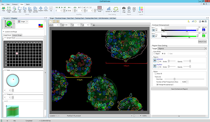
Yokogawa Cell Pathfinder (CPf)
Overview
Designed with both screeners and microscopists in mind, CPf utilizes simple application templates for standard assays such as Cell Cycle and Translocation, a simplified basic mode for users who are new to high content analysis, and an advanced mode with over 100 selections to optimize object identification.
All modes are designed with a flexible and straightforward workflow-based interface that makes the entire process simple but powerful.
Cell Pathfinder Features
- Live Cell Long Term Time Lapse: image a plate for 3+ days while maintaining healthy cells utilizing the CV8000’s CO2, Temperature and Active Humidification
- CPf’s Cell Tracking module provides 9 parameters users can adjust to ensure the same object is consistently tracked frame to frame
- Track cell division. Output links mother cells to daughter cells
- Track speed, distance moved, Linearity of Motion
- Live Cell High Speed Time Lapse image at almost 50fps and add inhibitors/activators in real time using the CV8000’s built in disposable tip pipettor
- CPf’s Time in Same Region model creates a maximum image projection across time. This enables CPf to create a single fixed area to measure changes over the course of a high speed time lapse assay.
- Run Ca+ assays, cardiomyocyte assays, and more
- 3D Imaging: build 3D models of your cells, pull out more information, and expand the knowledge of your biological system
- Report volume, surface area, and count objects within spheroids, organoids, or PDX tumor models.
- Analyze in 3D or convert to maximum, minimum, average, or sum image projection
- SearchFirst™ : quickly capture rare events, improve imaging speed, and locate spheroids
- First Scan using low powered objectives then acquire objects of interest at high resolution
- Machine Learning and Manual Controls : Utilize the machine learning module to teach CPf how to distinguish objects of interest from each other and from background.
- Extremely powerful tool for ill-defined objects
- Manually trace objects of interest or regions of interest to ensure only those objects important to the experiment are being captured.
- Texture Analysis : utilize texture analysis to identify differences between cell states that may not be apparent when using standard intensity-based output.
- Report Peak, Valley, Hole, Edge, Ridge, and Saddle textures
- Control pixel distance, test and compare results
- Gating : separate cell with distinct characteristics into populations using the gating module